Supporting patients and families with 3D modelling and VR
Medical and research teams at Great Ormond Street Hospital for Children (GOSH) in London are using technology to help children and families prepare for surgery. Joe, aged 15, is being treated at GOSH for Marfan Syndrome, a genetic disorder that affects the body’s connective tissue, which plays an important role in helping the body grow and develop properly. Joe has an associated heart condition and required surgery to replace his aorta, the body’s main artery.
His cardiac team share with us how they used pioneering 3D Heart Modelling and Virtual Reality to help Joe, his family and the surgical team understand his heart condition and prepare for his surgery.
Virtual reality (VR) in research
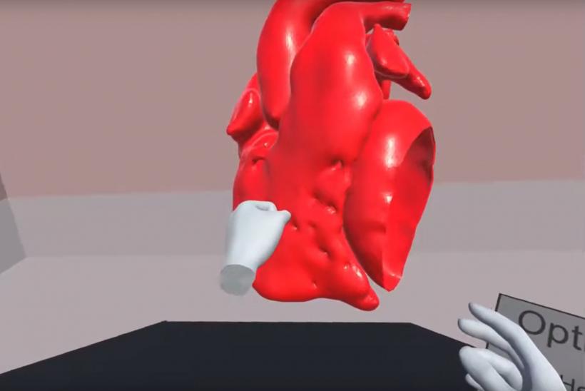
Research played a key part in the preparation for Joe’s cardiac surgery, with a 3D model image of his heart being captured and then looked at via Virtual Reality software. Dr Claudio Capelli and Endrit Pajaziti, research fellows based at GOSH, explain the process:
“When a patient has a CT or MRI scan we take the images and then transform these into a 3D model of the heart. This 3D model can be then be 3D printed or imported in our VR software, which was designed and developed in house by our team.
“Our group have published several studies showing how 3D printed models can be beneficial to all the people involved in the care of our patients. Our current research focuses on the long-term benefit that young teenagers, such as Joe, might have in engaging with their heart condition also by means of 3D printed models.”
How VR and 3D modelling helped Joe
Claudio explains: “VR is a fantastic way of being immersed into a patient’s heart. The overall aim of using VR in our research projects is to help us to explore the cardiac anatomy in an immersive way, providing more detail than ever before.
“Firstly, it can help the clinicians during their decision phase. A VR model can enhance the understanding of a patient’s anatomy so that the clinical team can plan a surgical procedure in the virtual world prior to theatre. The surgeon can see the heart from any perspective (including the inside), take detailed measurements and virtually plan and practice procedures.
“Secondly, we aim to improve the education of congenital heart disease. In this case, we are building a virtual library of models of the most common congenital heart diseases for medical students and young trainees, to help them understand more about the condition.
“Finally, we believe that VR can be a tool to engage with our patients. For Joe, VR not only stimulated curiosity in understanding his condition, but helped in entertaining and distracting him before his surgery.”
Elena Cervi, Joe’s Cardiology Consultant at GOSH, adds “3D models are proving very useful to explain diseases and procedures in a more visual and understandable way to patients and parents. They are usually thrilled to keep the model (when it is 3D printed) as it is made from their own imaging. Unlike a standard prop for teaching this is the anatomy specific to the single patient which makes it unique.”
The VR project at GOSH has been initially funded by La Fondation Dassault Systemes. The 3D printed model research projects have been funded by British Heart Foundation.

